A mid-50s female presents with 3 days of shortness of breath, abdominal pain, and vomiting. Initial Vitals: Temp 98.8 ℉, HR 112, BP 84/52, RR 22, SpO₂ 96% on RA.
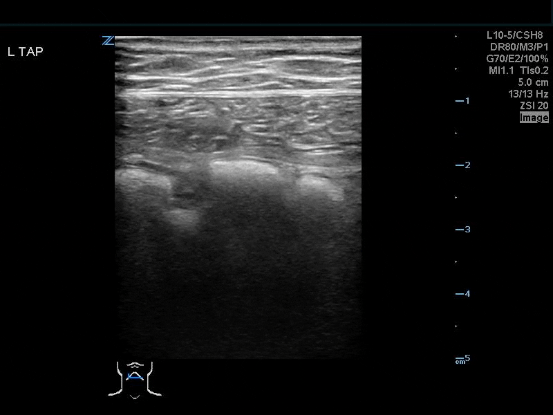
Given her undifferentiated shock you perform a RUSH Exam, which is otherwise unremarkable with the exception of the following clip:
Your interpretation? Anechoic appearing area adjacent to the caudal liver tip. Bingo! Free fluid! You then order a CT of the ABD/PEL and…

It's normal...welp...that's #awkward
You found the tip like Brian & Bob always harp on...what happened?!
Lipliner Artifact!
The images from this real case demonstrate a characteristic example of what has been recently termed as the “Lipliner Sign”!
In December of 2024, a prominent article was published in JEM after clinicians within the ultrasound community noticed a trend of similar ultrasound findings that resulted in false positive FAST exams. And what did they find? It was our own ultrasound machines that was the culprit! Essentially, the technology of modern ultrasound machines does a really good job at enhancing overall image quality in real-time as you scan (through real-time adaptive filtering to reduce noise/speckling and increase spatial resolution, for you ultrasound physics nerds out there). While these advanced imaging processing technologies are most often beneficial, they have also been found to cause a “lipliner artifact”, that can result in false positive FAST exams.
So now, the major clinical question: Lipliner Artifact vs. Free-Fluid?!
Lipliner sign/artifact is a simple, linear, anechoic feature that outlines the adjacent solid organ. It is predominantly found at the edges of solid organs (i.e. liver and spleen), which unfortunately are the same locations where free-fluid is likely to be found on FAST exams.

In contrast, free-fluid forms a wedge shape that decreases in width as it dissects dependently into tissue planes.

Well, what can you do then to differentiate between lipliner sign & free fluid?!
Rotate your probe and change to an orthogonal imaging plane!
If the anechoic area of concern significantly decreases or disappears, then likely lipliner artifact. If it remains, then suspect free-fluid.
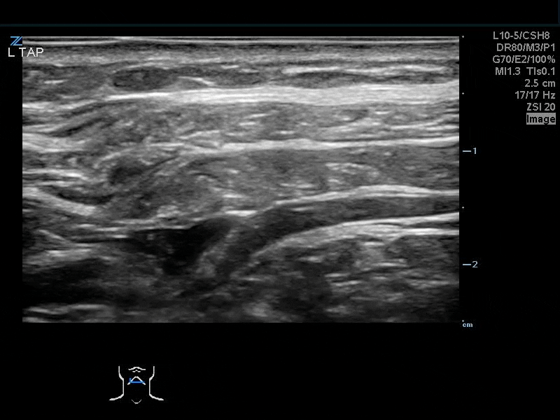
Change the machine settings!
On Mindray machines, iClear is the proprietary feature that reduces image-adpative artifact.
Under the “Image” tab, you can reduce the iClear settings, or even turn it off to 0 if needed
See below on where the iClear setting is located on our TEX machines!

SUMMARY
Lipliner Sign is a machine processing artifact that can be found along the edges of solid organs, resulting false positive FAST exams
Free Fluid often takes a more wedge-shaped appearance, and tapers at the edges as it dissects dependently into tissues
Attempts to differentiate between Lipliner & Free-Fluid can be performed by:
Rotating probe to an orthogonal plane
Decreasing/turn off the iClear setting
References:
Lipliner Artifact Review. Acep.org. Published 2025. Accessed May 11, 2026. https://www.acep.org/emultrasound/newsroom/march-2025/lipliner-artifact-review
Parker MA, Hicks BG, Kaili M, et al. The Lipliner Sign: Potential Cause of a False Positive Focused Assessment with Sonography in Trauma (FAST) Examination. The Journal of Emergency Medicine. 2024;67(6):e553-e559. doi:https://doi.org/10.1016/j.jemermed.2024.06.013
Stolz L, Ferre R. Managing and Conquering the Lipliner Artifact. The Journal of Emergency Medicine. 2025;76:157-158. doi:https://doi.org/10.1016/j.jemermed.2025.04.034